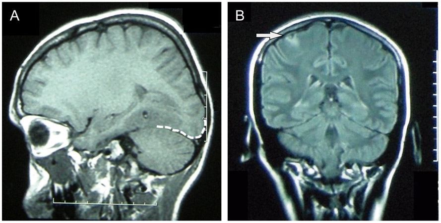
Fig. 1.
MRI images of two representative patients. (A) T1-weighted MRI of a cysticercotic viable cyst localized in a neocortical temporal area showing perilesional edema that reached the occipital lobe (approximation of the occipital lobe cortex demarcated with a white dashed line). The patient had multiple visual seizures from 60 days to seven days before the acquisition of this MRI. (B) Fluid-attenuated inversion recovery (FLAIR) MRI image showing a viable cyst in the right perirolandic area (arrow). This patient experienced a tonic seizure of the left arm and the left-sided face, as she had presented previously, during an EEG that was showing right fronto-central polyspikes at that time.

