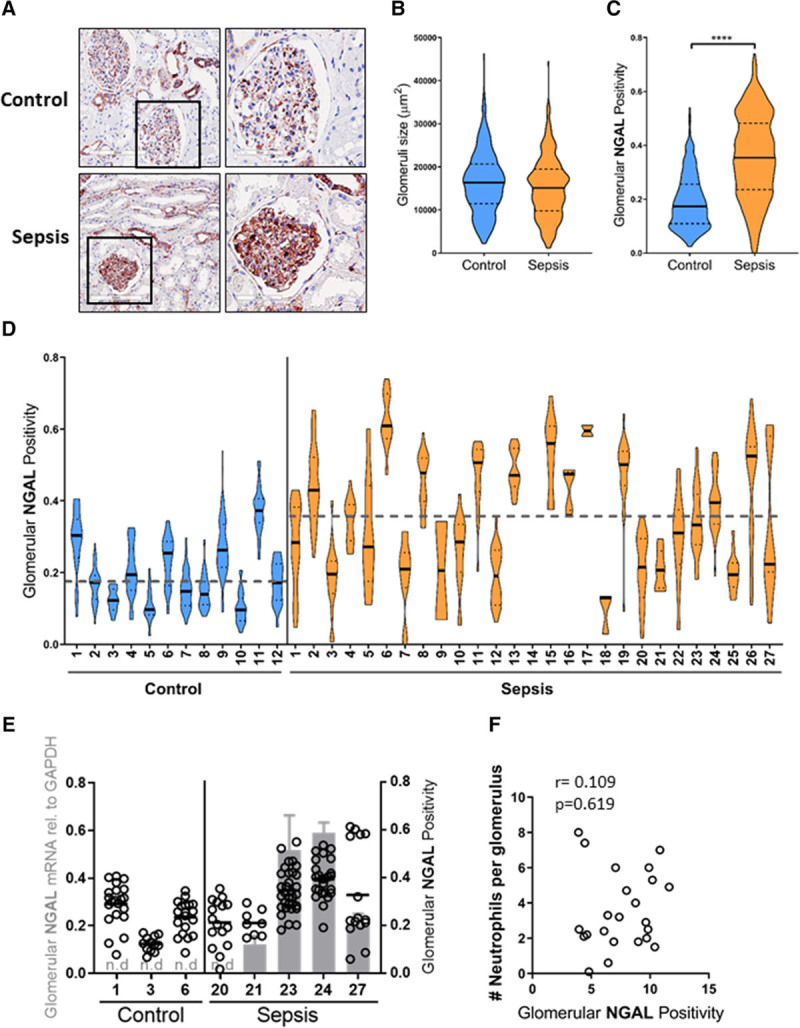
Figure 3.

Glomerular neutrophil gelatinase–associated lipocalin (NGAL) protein levels identifies glomerular heterogeneity in sepsis-acute kidney injury (AKI) patients. A, Representative immunohistochemical staining of NGAL (red) in a postmortem kidney biopsy from a sepsis-AKI patient compared with control renal tissue, original magnification ×400. B, Violin plot of glomerulus size of all individual glomeruli per biopsy in control (n = 469 from 12 control biopsies) and sepsis-AKI patients (n = 406 from 27 sepsis-AKI biopsies). The solid lines indicate the median size of all glomeruli in control and sepsis-AKI subjects, respectively. C, Violin plot of glomerular NGAL positivity in all glomeruli tufts from all control (n = 12) and sepsis-AKI patients (n = 27). The solid lines indicate the median glomerular positivity in control and sepsis-AKI subjects, respectively. D, Glomerular NGAL positivity was determined in all glomeruli tufts within a renal biopsy from control subjects (n = 12) and sepsis-AKI (n = 27). Each dot represents the NGAL positivity within a single glomerulus within a single renal biopsy. Solid lines indicate the median glomerular positivity in that particular biopsy. The dashed lines indicate the mean size of all glomeruli in control and sepsis-AKI subjects, respectively. E, Glomerular NGAL messenger RNA levels were determined within the whole biopsy by reverse transcription quantitative polymerase chain reaction in control (n = 3) and sepsis-AKI patients (n = 5) using glyceraldehyde-3-phosphate dehydrogenase (GAPDH) as a housekeeping gene (gray bars, left axis). Dots indicate the individual glomerular NGAL positivity from the same control and sepsis-AKI patient (right axis). F, Glomerular NGAL protein levels determined in biopsies from sepsis-AKI patients do not correlate with the average number of neutrophils infiltrating glomeruli in the same sepsis-AKI patients. r = 0.109, p = 0.619.
