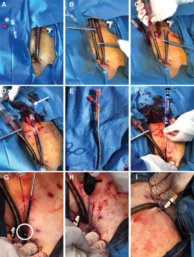
Figure 1.

Sterile preparation of the cannulation area with asterisk indicating hemostasis valve Y connector (A). Clamping of arterial and venous cannula shortly behind the selectively hardened proximal venous and arterial cannula body (B) and subsequent cutting by scissors (C). Insertion of hemostasis valve Y connector (Merit Angioplasty Pack, South Jordan, UT) (D) and wire insertion (arrow) into the proximal cannula (E). Insertion and releasing of the first ProGlide via guidewire (Abbott Vascular, Lake Bluff, IL) (F). Reinsertion of the guidewire into the side hole of the first ProGlide device (circle) to place a second ProGlide device (G). Tightening of knots by knot pusher (H). Preparation for removal of the venous cannula by insertion of a Z-suture (I).
