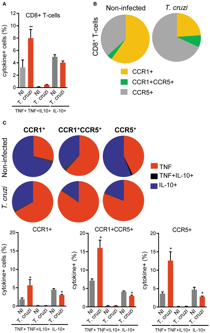
Figure 7.
Expression of TNF, IL-10 and CC-chemokine receptors CCR1 and CCR5 by CD8+ T-cells in spleen of T. cruzi-infected C57BL/6 mice. Mice were infected with 100 trypomastigote forms of the Colombian T. cruzi strain and analyzed at 120 days postinfection. Splenocytes were collected and stained for cell surface markers and intracellular cytokines (CD8, CCR1, CCR5; TNF, IL-10). After selection of singlets (FSC-Lin × FSC-Area, R1), dead-cell exclusion (FSC-A × SSC-Lin, R2), TCR × CD8 dot plot (gating on TCR+CD8+ cells, R3), TNF × IL-10 dot plots were analyzed. (A) Graph shows the frequencies of TNF+, TNF+ IL-10+, and IL-10+ cells among the CD8+ T-cells. (B) Pie charts represent the fractions of CD8+ T-cells obtained from spleens of NI and T. cruzi-infected mice that are CCR1+, CCR1+ CCR5+, and CCR5+, as indicated in the legend. (C) Pie charts represent the fractions of CCR1+, CCR1+ CCR5+, and CCR5+ CD8+ T-cells that carry each of the intracellular cytokine phenotypes shown in the legend. Data represent two independent experiments with three NI and four to five infected mice. *P < 0.05, **P < 0.01, NI vs. T. cruzi (ANOVA Bonferroni posttest).

