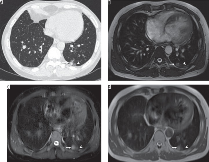
Figure 1.
A) HRCT, B) TRUFI, C) SPAIR, and D) HASTE – axial images of a 30-year-old male patient with lymphoma, on chemotherapy, complaining of cough and fever. Computed tomography image shows a well-defined nodule (arrow) with surrounding ground-glass opacities (arrowhead) in the posterobasal segment of the left lower lobe, which is also well demonstrated on magnetic resonance images

