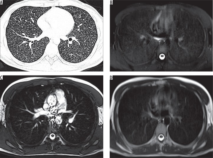
Figure 2.
A) HRCT, B) SPAIR, C) TRUFI, and D) BLADE – axial images of a 26-year-old male patient known case of lymphoma, on chemotherapy, complaining of cough and fever. Computed tomography shows miliary nodules in bilateral lungs. SPAIR magnetic resonance images show diffusely increased signal intensity with no discrete nodules. On TRUFI and BLADE, no abnormality is demonstrated

