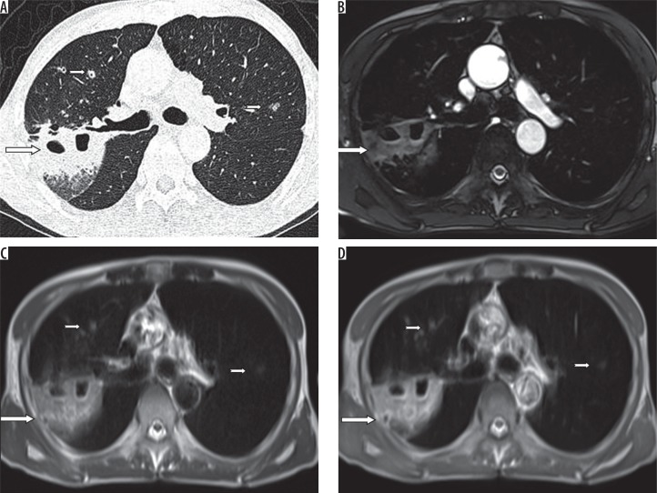
Figure 3.
A) HRCT, B) TRUFI, C) BLADE, and D) HASTE – axial images of a 48-year-old female patient of carcinoma ovary, on chemotherapy, complaining of cough and fever. Computed tomography shows a patchy area of consolidation (long arrows) with breakdown in the posterior segment of the right upper lobe and few nodules (short arrows) in the anterior segment of right upper lobe and the apicoposterior segment of left upper lobe, which are also well demonstrated on BLADE and HASTE images. On TRUFI sequence, nodules in the left upper lobe and right upper lobe are not demonstrated

