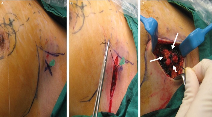
Figure 2.

A, The clipped node was localized by placing a 21 G needle perpendicular to skin at the cross marking. B, After raising the skin flaps, the needle was removed after a stitch had been placed at the needle site. C, With the stitch as the center, a 1 cm all‐round margin was marked out with blue ink (arrows) and resection along the blue ink performed
