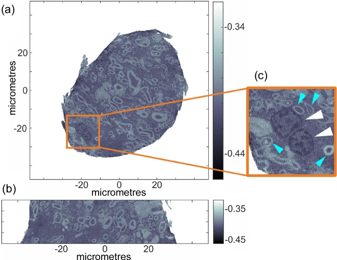
Figure 3.
Electron density tomogram of a cylindrical unstained brain tissue sample prepared using the lathe system under cryogenic conditions and measured using ptychographic X-ray computed tomography: (a) axial view; (b) sagittal view. Variations in grayscale represent changes in electron density (e A−3). (c) Magnified region of (a) showing nuclei with euchromatin (white arrowheads) and axonal cross-sections (blue arrowheads). PXCT measurements were conducted under cryogenic conditions.

