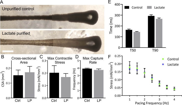
Fig 5. Lactate purified cardiomyocytes do not alter tissue formation via compaction and electromechanical function at one week.
(A) Engineered cardiac tissues made with 5% human cardiac fibroblasts (hCFs) and unpurified hiPSC-cardiomyocytes (38% cTnT+; top) or lactate purified hiPSC-cardiomyocytes (82% cTnT+; bottom) were cultured for one week. Scale bar: 1 mm; images stitched together. (B-F) Control tissues (Ctrl, n = 8) and tissues with lactate purified cells (LP, n = 4) underwent mechanics testing at 1 week. Cross-sectional area (B), maximum twitch contraction stress (C), and maximum capture rate (D) were measured. (E) Relaxation time to 50% (T50) and 90% (T90) of maximum stress was measured. (F) Maximum stress production was measured at increasing pacing frequency. Data are represented as mean ± SEM.

