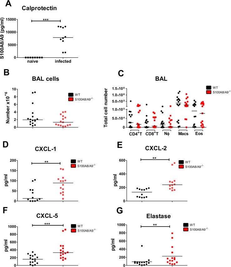
Fig 3. Increased immune responses to migrating L. sigmodontis L3 larvae within the BAL of S100A8/A9-/- mice.
Concentration of calprotectin (A) and total number of bronchoalveolar cells (B) as well as CD4+ T cell (CD4+ T), CD8+ T cell (CD8+ T), neutrophil (NØ), macrophage (Macs), and eosinophil (Eos) cell counts (C) in WT and S100A8/A9-/- mice 12 days after subcutaneous L. sigmodontis infection. Concentrations of CXCL-1 (D), CXCL-2 (E), CXCL-5 (F), and elastase (G) in the bronchoalveolar lavage 12 days after L. sigmodontis infection of WT and S100A8/A9-/- mice. Results are shown as median (A-G). Statistical significance was analyzed by two-tailed Mann-Whitney-U-test (A-G). **p<0.01., ***p<0.001. Data shown in A-G are pooled of 2–3 independent experiments with 5–8 mice per group.

