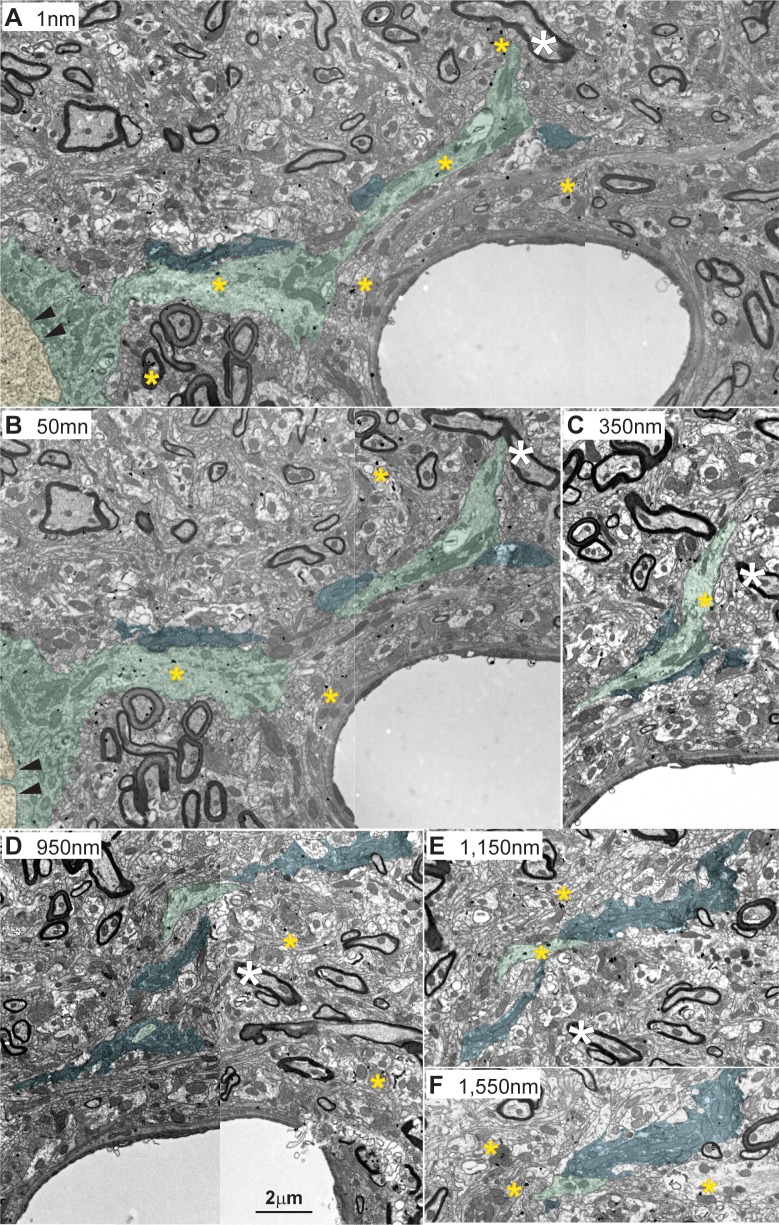
Fig 12. Amygdalar pathways form synapses proximal to neuron cell bodies in the MDmc.
(A–F) A series of consecutive sections captured from the electron microscope revealed that amygdala terminals (black label with blue overlay) wrapped around EDs (green) close to the neuron body, identified by the presence of the cellular nucleus (A and B; black double arrowhead and yellow). Amygdalar terminals were labeled with the neural tracer FE and visualized with DAB. Yellow asterisks show silver-enhanced gold labeling for PV in postsynaptic dendrites. Numbers in A–F indicate distance in nanometers between sections photographed. White asterisks show fiduciary marks. Calibration bar in D applies to A–F. DAB, diaminobenzidine; ED, excitatory dendrite; FE, Fluoro-emerald; MDmc, mediodorsal thalamic nucleus, magnocellular part; PV, parvalbumin.

