. 2020 Feb 13;128(2):027005. doi: 10.1289/EHP4709

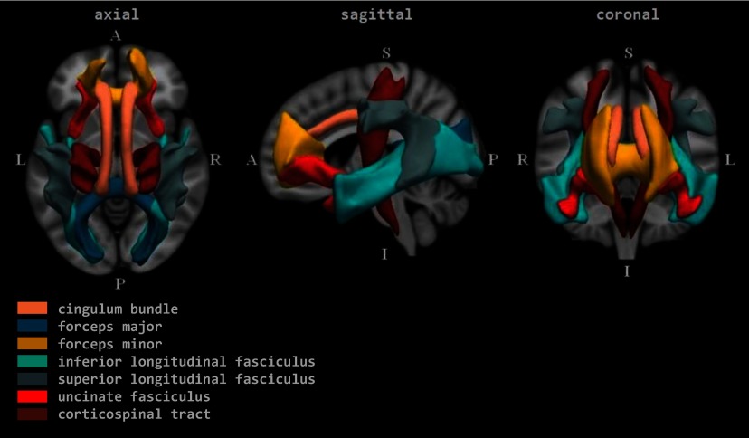
Figure 1.

Figure 1 comprises the axial, sagittal, and coronal views of the brain. In the axial, the anterior (at the top) has forceps minor, uncinate fasciculus, and a part of cingulum bundle; the left and right each have inferior longitudinal fasciculus, superior longitudinal fasciculus, forceps major, corticospinal tract, and parts of cingulum bundle; the posterior (at the bottom) has forceps major and inferior longitudinal fasciculus. In the sagittal, the superior (at the top) has parts of the corticospinal tract, cingulum bundle, and superior longitudinal fasciculus; the anterior (on the left) has forceps minor, uncinate fasciculus, a part of the cingulum bundle, and part of inferior longitudinal fasciculus; the posterior (on the right) has forceps major, inferior longitudinal fasciculus, superior longitudinal fasciculus, and a part of the corticospinal tract; the inferior (at the bottom) has the corticospinal tract, uncinate fasciculus, and inferior longitudinal fasciculus. In the coronal, the superior (at the top) has corticospinal tract, parts of cingulum bundle, forceps minor, and superior longitudinal fasciculus; the right and left each have corticospinal tract, superior longitudinal fasciculus, inferior longitudinal fasciculus, cingulum bundle, uncinate fasciculus, and forceps minor; the inferior (at the bottom) has parts of uncinate fasciculus, corticospinal tract, inferior longitudinal fasciculus, and forceps minor.

Group average representations of the tracts in standard coordinate space. Note: A, anterior; I, inferior; L, left; P, posterior; R, right; S, superior.