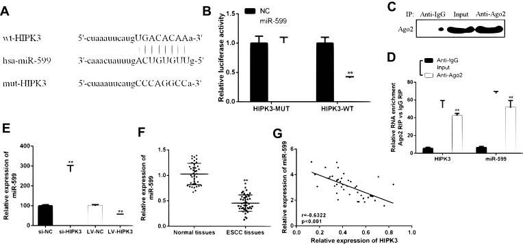
Figure 3.
HIPK3 directly targeted miR-599 in ESCC. (A) Bioinformatics analysis revealed predicted binding sites between HIPK3 and miR-599. (B) Relative luciferase activity in TE-13 cells co-transfected with wild-type (HIPK3-WT) or mutant reporter plasmid (HIPK3-mut) and miR-599 mimic or negative control. (C) Cellular lysates from ESCC cells were used for RIP with an Ago2 antibody. The Ago2 protein level was detected by Western blot, (D) and the relative expression of HIPK3 and miR-599 in the immunoprecipitate was measured by RT-PCR. (E) The miR-599 expression in ESCC tissues and matched normal tissues. (F) Expression of miR-599 in the presence of si-HIPK3 or LV-HIPK3. (G) The relationship between HIPK3 and miR-599 in the ESCC organization.(** P <0.01).

