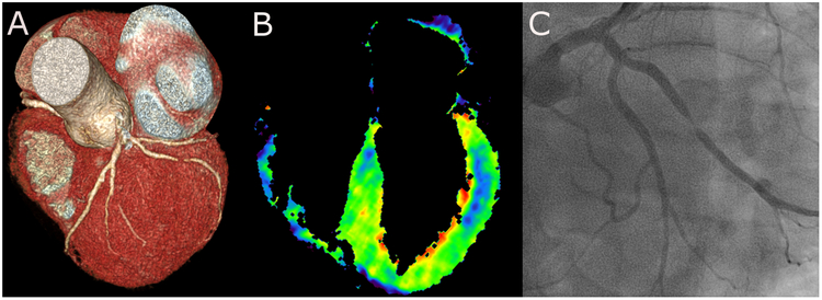
Figure 1: Acquisition protocol.
For a “rest-stress approach”, CTA is performed first, followed by dynamic stress CT myocardial perfusion imaging in case of abnormalities. A delay between both scans is required for contrast medium washout. A low-dose native scan is performed during systole to plan the perfusion scan, which is also performed during systole. Attenuation values throughout the myocardium and the aorta are plotted against time, from which myocardial perfusion maps can be reconstructed. Nitroglycerin (NTG), beta-blocker (BB), contrast medium (CM), myocardial perfusion imaging (MPI), myocardial blood flow (MBF).

