Figure 2.
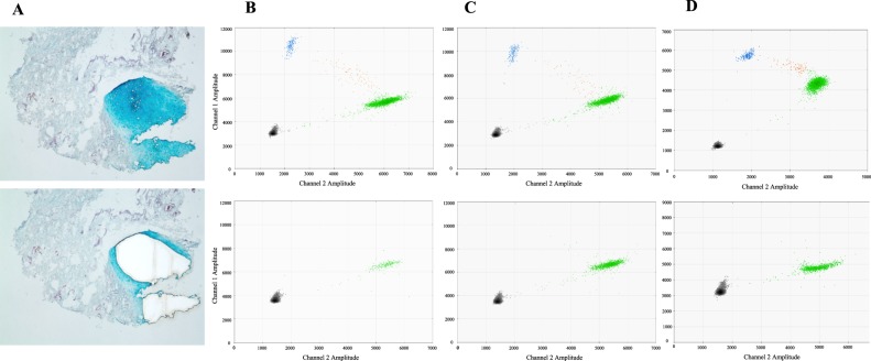
Somatic MAP2K1 mutations are isolated to the skin and subcutis of ear AVM tissue. (A) Laser capture microdissection of cartilage from surrounding tissue to minimize inclusion of adjacent microscopic vessels containing mutant endothelial cells (Alcian Blue stain; Patient 2). Top panel = pre-microdissection, bottom panel = post-microdissection. B,C,D = Patient 1, 2, 3 ddPCR graphs of their AVM ear tissue. Top row of graphs = skin and subcutaneous adipose. Bottom row of graphs = cartilage. Left upper blue droplets contain mutant alleles. Right middle orange droplets have mutant and wild-type alleles. Right lower green droplets contain wild-type alleles. Left lower black droplets are empty. Note absence of mutant droplets in the cartilage graphs.

