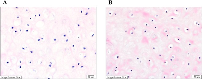
Figure 3.
Histological appearance of overgrown AVM cartilage and normal cartilage is similar. Sections of (A) conchal ear cartilage from a patient with an AVM (Patient 3). (B) Control conchal ear cartilage from a patient with a normal ear. Sections show equivalent distribution and cellularity of chondrocytes in a chondromyxoid matrix. The chondrocytes have normal appearance with monomorphic pyknotic nuclei. (Hematoxylin and eosin stain, 20x magnification, scale bar 20 µm).

