Figure 3.
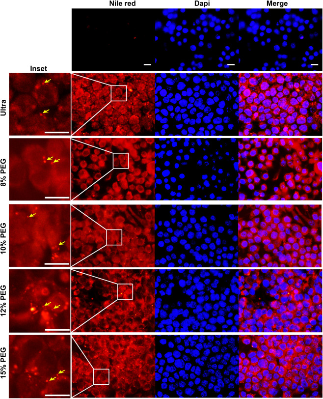
Intracellular uptake of PEG-ENPs is similar to ultra-ENPs in murine macrophages. RAW cells were either mock treated or treated with Nile red labeled ultra or PEG-ENPs (100 µg), for 24 hours. Cells were fixed and counterstained with the nuclear stain, DAPI. Top panel shows mock treated cells with no detectable fluorescence. Cytoplasmic Nile Red fluorescence was detected in all ENP treated cells indicating the intra-cellular uptake of both ultra and PEG-ENPs by murine macrophages (bottom panels). No significance difference in fluorescence intensity or the number of Nile red positive cells was observed between these two different treatments. Inset shows amplified regions depicted in the box to demonstrate the presence of Nile red labeled nano-sized ENPs. Scale bar-10 µm.

