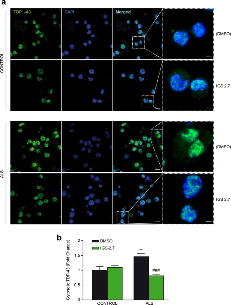
Figure 5.
Subcellular localization of TDP-43 after IGS-2.7 treatment of lymphoblasts from control and ALS subjects. Lymphoblasts were seeded at 106 cells × ml−1 and incubated in presence or absence of IGS-2.7 (5 μM) for 24 h. (a) Cells were stained with anti-TDP-43 antibody followed by a secondary antibody labeled with Alexa Fluor 488. DAPI was included in the mounting media to stain the nucleus. TDP-43 protein localization was assessed by confocal laser scanning microscopy. Merged images show that treatment with IGS-2.7 prevented the higher cytosolic accumulation (red arrows) of TDP-43 protein in ALS patients. Scale bars = 11 μM. Magnified cells from images are shown on the right panels for better visualization (Scale bars = 3 μm) (b) Quantification of TDP-43 cytosolic localization in lymphoblasts from ALS patients compared to controls. Data are expressed as mean ± SEM for experiments carried out with 4 different cell lines for each group. Data were assessed by one-way ANOVA and post hoc Fisher’s analysis (**p < 0.01 from control cells; ###p < 0.001 from untreated ALS cells).

