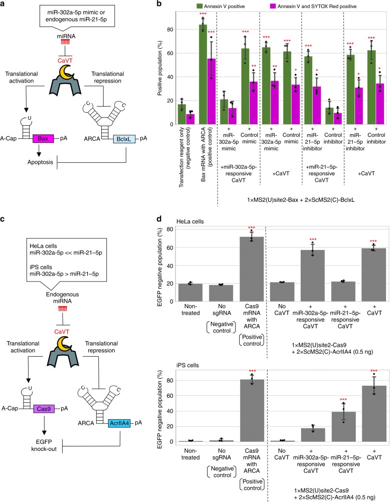
Fig. 7. miRNA-mediated simultaneous regulation of translational activation and repression using miRNA-responsive CaVT mRNA.
a, b Apoptosis-inducing circuit regulated by miRNA. HeLa cells were co-transfected with 1xMS2(U)site2-Bax mRNA (cap analog: A-cap), 2xScMS2(C)-BclxL mRNA (cap analog: ARCA), the indicated CaVT mRNA, and the indicated miRNA mimic or inhibitor. For the positive control, 1xMS2(U)site2-Bax mRNA (cap analog: ARCA) was transfected. All mRNAs contained N1mΨ. One day after the transfection, the cells were stained with Annexin V and SYTOX Red followed by flow cytometry. A schematic diagram of the RNA circuit (a). The bar graph shows the average of three independent experiments (mean ± SD) (b). *P < 0.05, **P < 0.01, ***P < 0.001 compared to the negative control by ANOVA with Dunnett’s multiple comparison test (two-sided). Exact P values are shown in Supplementary Table 1. Source data are provided as a Source Data file. c, d Cell-selective regulation of EGFP knockout by a miRNA-responsive genome editing circuit. The indicated EGFP-expressing stable cell lines were co-transfected with 1xMS2(U)site2-SpCas9 mRNA (cap analog: A-cap), 2xScMS2(C)-AcrIIA4 mRNA (cap analog: ARCA), the indicated CaVT mRNA, and EGFP-targeting sgRNA. For the positive and negative control, 1xMS2(U)site2-SpCas9 (cap analog: ARCA) with or without EGFP-targeting sgRNA was transfected, respectively. All mRNAs contained N1mΨ. Five days after the transfection, EGFP fluorescence was analyzed by a flow cytometer. A schematic diagram of the RNA circuit (c). The bar graph shows the average of three independent experiments (mean ± SD) (d). ***P < 0.001 compared to the non-treated samples by ANOVA with Dunnett’s multiple comparison test (two-sided). Exact P values are shown in Supplementary Table 1. Source data are provided as a Source Data file.

