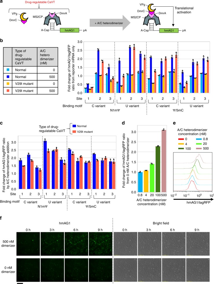
Fig. 8. Drug-mediated translational activation combining CaVT and the hetero-dimerization system.
HeLa cells were co-transfected with hmAG1 mRNAs containing an MS2-binding motif (cap analog: A-cap, modified nucleosides: N1mΨ or Ψ/5mC) and mRNAs that express tagRFP, DmrC-VPg(FCV), and MS2CP-1xDmrA or MS2CP(V29I)-1xDmrA. Then, A/C heterodimerizer was added. a Schematic diagram of drug-regulatable CaVT, which is composed of MS2CP-1xDmrA and DmrC-VPg(FCV). MS2CP-1xDmrA binds to the MS2 binding motif of target mRNAs. In the absence of A/C heterodimerizer, DmrC-VPg does not bind to DmrA, and there is no VPg-mediated translational activation. After the addition of A/C heterodimerizer, DmrA-DmrC interaction tethers VPg to the target mRNAs, and VPg activates translation. b, c Effects of modified nucleosides, sites, and variants of the MS2-binding motif and variants of MS2CP on translation. The fluorescence was measured by flow cytometry and means of the hmAG1/tagRFP ratio were normalized by the ratio in the reporter mRNA only (b) or 0 nM A/C heterodimerizer samples (c). The bar graph shows the average of three independent experiments (mean ± SD). Source data are provided as a Source Data file. d, e Dose-dependency of drug-regulatable CaVT. 1xMS2(U)site1-hmAG1 (cap analog: A-cap, modified nucleosides: N1mΨ), tagRFP, DmrC-VPg(FCV), and MS2CP-1xDmrA mRNAs were used for the transfection. Means of the hmAG1/tagRFP ratio were normalized by the ratio in 0 nM A/C heterodimerizer samples. The bar graph shows the average of three independent experiments (mean ± SD) (d). A representative histogram (e). Source data are provided as a Source Data file. f Time lapse images of drug-induced translational activation. 1xMS2(U)site1-hmAG1 (cap analog: A-cap, modified nucleosides: N1mΨ), DmrC-VPg(FCV), and MS2CP-1xDmrA mRNAs were used for the transfection. Each image was captured at the indicated time points after the addition of A/C heterodimerizer. Three independent experiments were performed to check reproducibility, and representative images are shown. The scale bar represents 200 μm.

