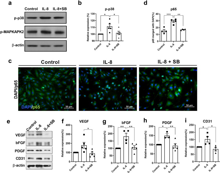
Figure 5.
Effects of p38 inhibition in vitro on IL-8-mediated angiogenic growth factor expression in mouse brain vascular bEnd.3 cells. (a) A significant increase in p-p38 was noted after IL-8 treatment (50 ng/ml), while SB203580 (50 μM) application 30 min prior to IL-8 administration decreased p-MAPKAPK2 levels according to Western blot analysis of OGD-conditioned bEnd.3 cells. (b) The graph depicts the band intensity of p-p38 in the Western blot assay. Data are shown as the mean ± SEM (each n = 5). (c) The proportion of merged p65- and DAPI-stained cells was significantly greater at 6 h after IL-8 treatment than in the OGD-only control cells and p38-inhibited cells upon immunocytochemistry analysis (200×). (d) The graph depicts the proportion of merged p65 and DAPI-stained cells. Data are shown as the mean ± SEM (each n = 4). (e–i) Westerns blot analysis of cells harvested 12 h after IL-8 treatment showed the upregulation of VEGF, bFGF and PDGF and the endothelial marker CD31, whereas cells pretreated with SB203580 exhibited the downregulation of the angiogenic growth factors and CD31, as shown in the graphs depicting the band intensity. Data are shown as the mean ± SEM (each n ≥ 4). The data shown in the graphs were collected from triplicate results. Asterisks (*) indicate significant differences (*p < 0.05, **p < 0.01, one-way ANOVA) according to Tukey’s multiple comparisons between groups. OGD, oxygen-glucose deprivation; Green, p65; DAPI, 4′, 6-diamidino-2-phenylindole for nuclear staining; Control, bEnd.3 cells without IL-8 treatment; IL-8+SB, SB203580 infused prior to IL-8 administration.

