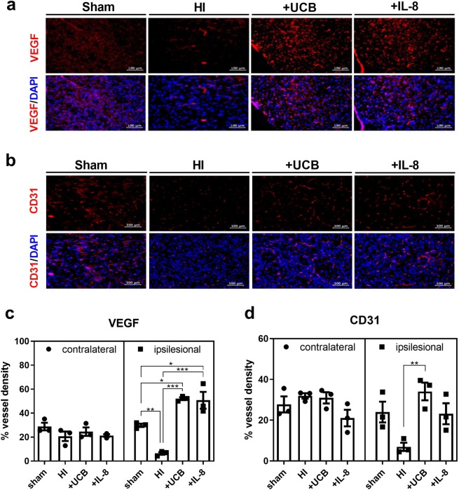
Figure 6.
Effects of hUCBCs and IL-8 on angiogenesis in vivo in the HI mouse brain. Angiogenesis was observed in vivo through immunohistochemistry staining for VEGF and CD31 in the peri-infarct cortex and striatum of HI mice dissected one week after either hUCBC (3 × 10⁷/kg) or IL-8 (50 µg/kg) injection. (a,c) The graph depicts the density of vessels (percentage) expressing VEGF among all cells stained with DAPI according to IHC analysis in the ipsilesional and contralateral hemispheres. (b,d) The graph depicts the percentage of vessel density expressing CD31 among DAPI-stained cells observed in the lesion side and unaffected contralateral hemispheres. On the other hand, the unaffected contralateral brain hemisphere was not significantly affected by HI, hUCBC or IL-8 administration. IHC findings of the contralateral hemisphere are depicted in Supplementary Fig. S3. Data are shown as the mean ± SEM (each n = 3). Asterisks (*) indicate significant differences (*p < 0.05, **p < 0.01, ***p < 0.001, one-way ANOVA) according to Tukey’s multiple comparisons between groups. HI, hypoxic-ischaemic brain injury; DAPI, 4′, 6-diamidino-2-phenylindole for nuclear staining; +UCB, HI mice subjected to hUCBC treatment; +IL-8, HI mice subjected to IL-8 treatment; hUCBCs, human umbilical cord blood mononuclear cells.

