FIG 9.
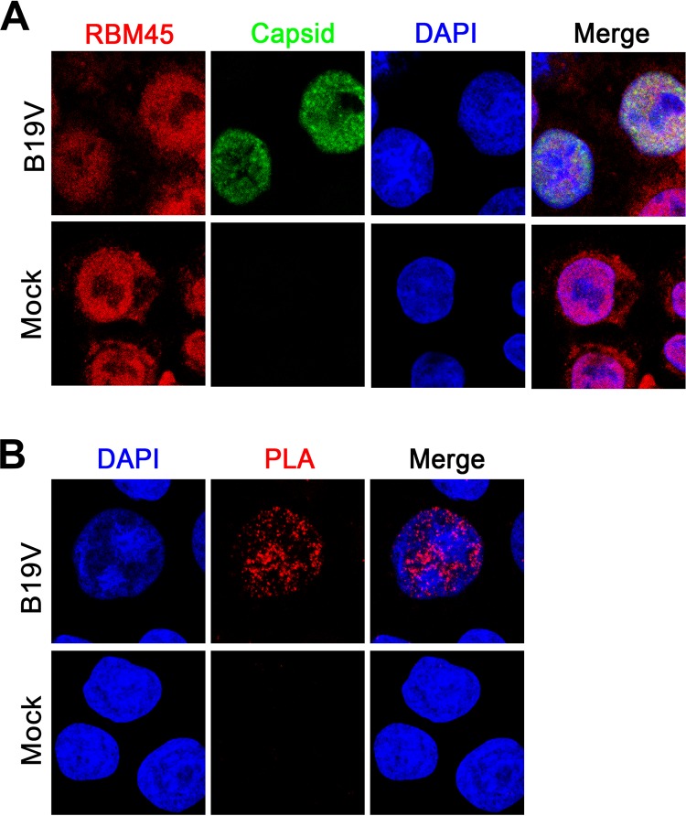
RBM45 was colocalized within the viral DNA replication centers indicated by capsid detection. CD36+ EPCs were mock infected or infected with B19V. At 24 h postinfection, the cells were analyzed by immunofluorescence assay. (A) Confocal colocalization. Mock-infected or B19V-infected CD36+ EPCs were costained and incubated with rabbit anti-RBM45 and mouse anti-B19V capsid antibodies followed by Alexa Fluor 594-conjugated and FITC-conjugated secondary antibody. (B) Proximity ligation assay (PLA). Infected cells were costained with rabbit anti-RBM45 and mouse anti-B19V capsid antibodies, followed by a proximity ligation assay, which detected two proteins detected by two labeled secondary antibodies in close proximity (∼40 nm apart). Images were taken with a Leica TCS SPE confocal microscope at ×63 magnification. Nuclei were stained with DAPI.

