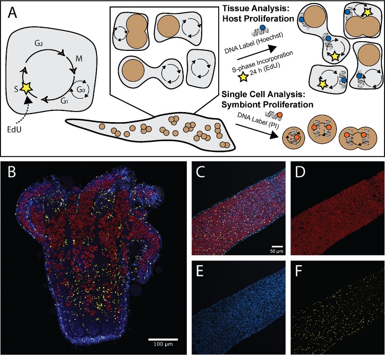
FIG 1.
Fluorescent labeling techniques used to correlate host nucleus proliferation and symbionts in Aiptasia-Symbiodiniaceae symbiosis. (A) Fluorescent labels were used to identify proliferating populations of host cells and cell cycle populations of symbiont cells. The presence of EdU incorporation over 24 h marked host cell proliferation, whereas all host nuclei were labeled with Hoechst stain. Symbionts were isolated from hosts or cultures and were labeled with propidium iodide to identify cell cycle populations based on DNA content. (B) Tiled section of a symbiotic Aiptasia sea anemone. Symbiont location was captured using chlorophyll autofluorescence shown in red. Cnidarian host nuclei were labeled with Hoechst stain (blue), and proliferating host nuclei were labeled with EdU-AF555 (yellow) to capture cell populations that have incorporated new DNA during S phase. (C to F) Host cell proliferation analyses were performed using fluorescently labeled Aiptasia tentacles (C) to allow for quantification and location mapping of symbionts (D), cnidarian host nuclei (E), and proliferating host nuclei (F).

