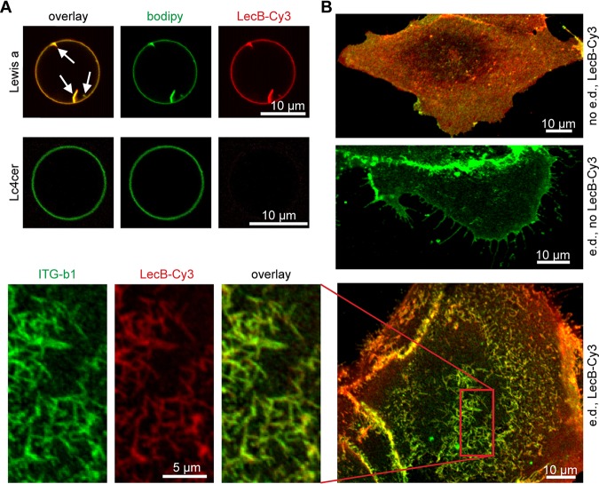
FIG 4.
Mechanism of LecB-mediated integrin internalization via cross-linking glycosphingolipids and integrins. (A) LecB-Cy3 (15 μg/ml, red) was applied to GUVs containing fucosylated glycosphingolipids bearing the Lewis a antigen (Lewis a) or the nonfucosylated precursor lactotetraosylceramide (Lc4cer) and BODIPY FL C5 HPC (bodipy; green) as a membrane marker. Confocal sections along equatorial planes of the GUVs are displayed; arrows point to membrane invaginations caused by LecB. (B) Subconfluent MDCK cells grown on glass coverslips were energy depleted (e.d.) or left untreated (no e.d.). LecB-Cy3 (red) was applied to the cells for 1 h, and cells were fixed and stained for β1-integrin (green). Confocal x-y sections at the level of the cell adhesion to the glass coverslip are displayed.

