
Figure 1.
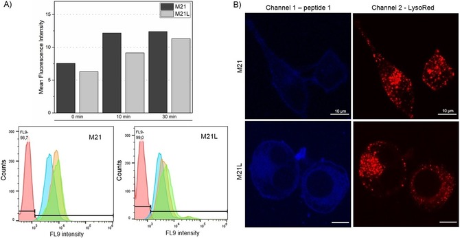
Uptake studies with peptide 1. A) Flow cytometry analysis after incubation of cells with peptide 1 (30 μm) for 0 min (blue), 10 min (orange), and 30 min (green). No peptide was added in the negative control (red). B) M21 and M21L cells were stained with LysoTracker Red DND‐99 and incubated for 10 min with peptide 1 (5 μm) prior to fluorescence microscopy. In combination both experiments clearly show an unspecific, fluid‐phase uptake of peptide 1.