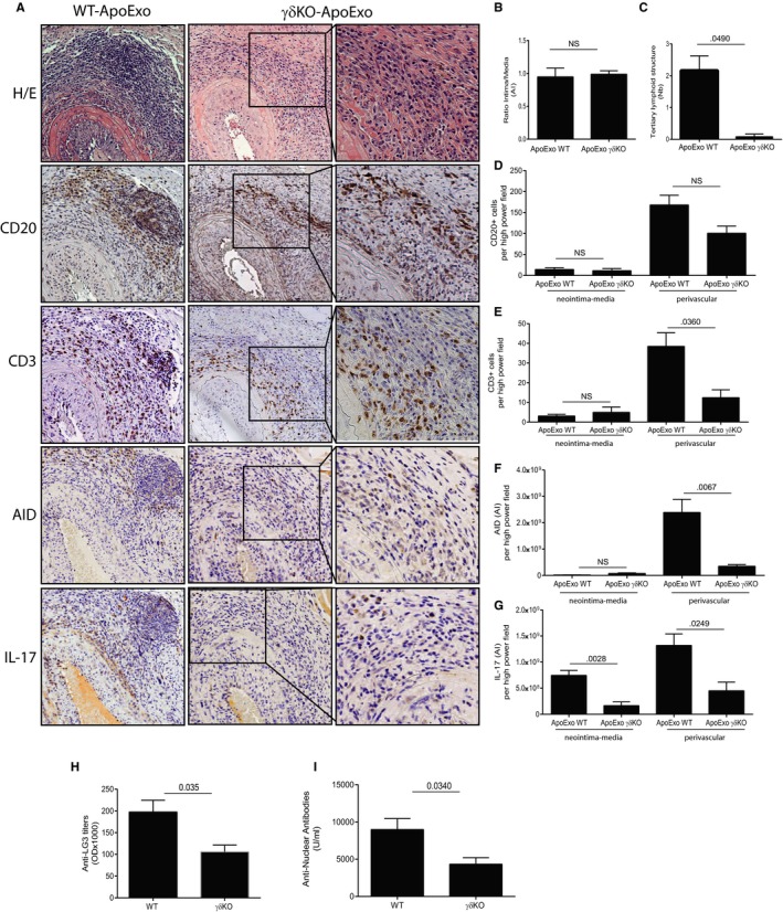
Figure 4.

Absence of γδT cells reduces allograft CD3+ T cell infiltration and IL‐17 expression, abrogates tertiary lymphoid structure (TLS) formation and reduces the formation of anti‐perlecan/LG3 and ANA. γδKO (n = 4) or wild‐type (n = 4) mice were injected with apoptotic exosome‐like vesicles (ApoExo) for 3 weeks posttransplantation: (A) Aortic allograft sections stained with H&E, CD20, CD3, AID, and IL‐17 (magnification: 5×; magnification of right inset panels: 20×). (B) Ratio intima/media in the allografts. (C) Mean number of TLS per allograft. Neointima‐media and perivascular quantification of CD20+ B cells (D), CD3+ T cells (E), AID (f), and IL‐17 (g) staining in each high‐power field of the allografts. Anti‐LG3 (h) and ANA (i) IgG levels in sera from γδKO or wild‐type allografted mice after 3 weeks of intravenous injections with ApoExo. Data were pooled from two independent experiments and expressed as means ± SEM. Comparison with the vehicle was done with a Student's t test
