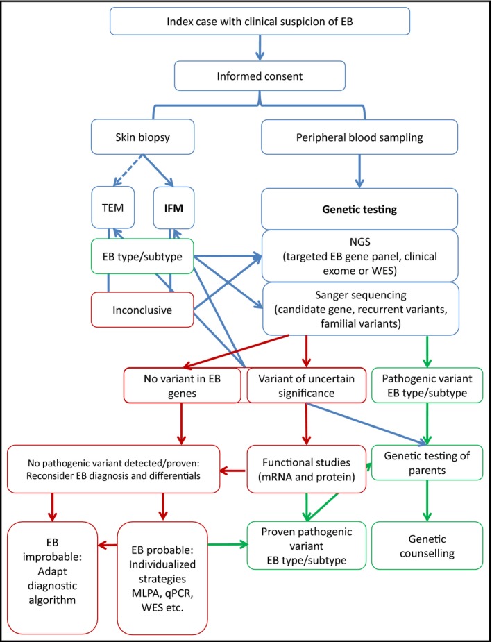
Figure 2.

Flowchart of laboratory diagnosis of epidermolysis bullosa (EB). Schematic representation of the steps required to achieve a molecular diagnosis of EB. Steps shown in green lead to a clear diagnosis of the EB type or subtype, while steps shown in red may require individualized strategies in a research setting. IFM, immunofluorescence mapping; MLPA, multiplex ligation‐dependent probe amplification; NGS, next‐generation sequencing; qPCR, quantitative polymerase chain reaction; TEM, transmission electron microscopy; WES, whole‐exome sequencing.
