Figure 1.

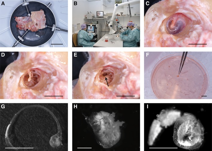
Tissue isolation from autopsy temporal bones. Unfixed human temporal bones (A) were obtained from the Pathology Department and dissected in the temporal bone laboratory (B). As a first step, the external ear canal is enlarged to allow visualization of the ear drum (C). After removal of the ear drum (D) and the ossicles (E), the inner ear was opened at the oval window (arrow in E) to remove the utricle and the membranous labyrinth, which are directly placed into cell culture medium (F). Further drilling was needed to remove the membranous part of the scala media (not shown). The membranous semicircular canal with the ampullary end (G), the utricle (H), and the membranous part of the cochlear scala media (I) are shown in higher magnification. Scale bars = 4 cm (A), 1 cm (C–F), 0.5 cm (G, I), and 0.2 cm (H).
