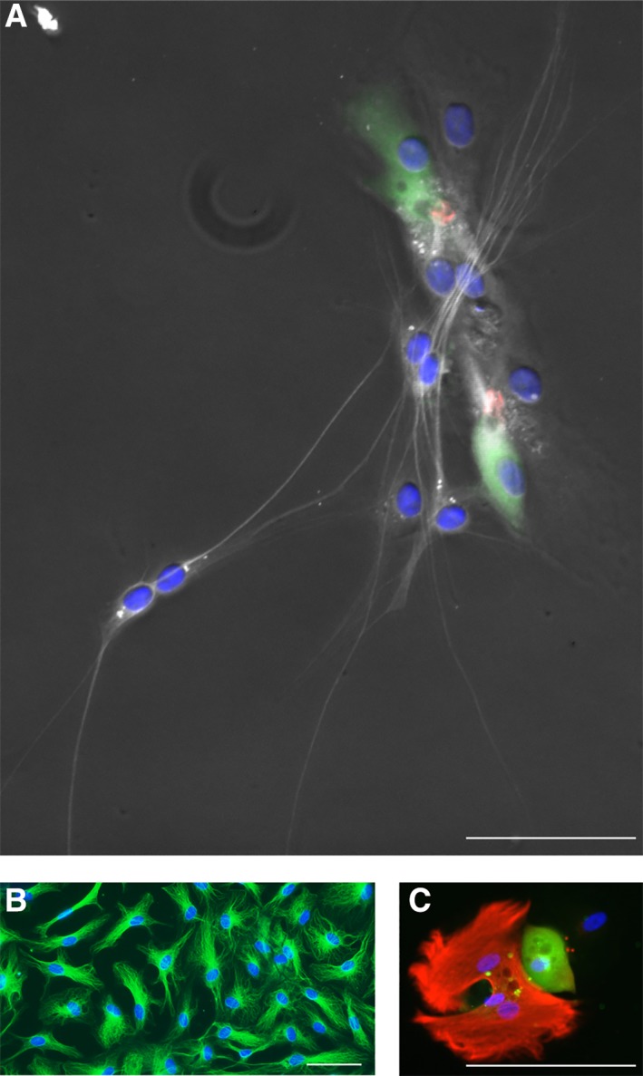
Figure 5.

Neuronal and glial cell types. Cell types with neuronal morphology are labeled with a marker for ß‐III tubulin (TUJ, white, A). These cells were generated from postmortem utricle‐derived spheres of a 72‐year‐old male donor (same patch of cells as in Fig. 4C). Homogeneous appearing cells with no neural morphology (TUJ, green, B) were obtained from cochlear duct‐derived spheres of an 80‐year‐old male postmortem donor. A glial cell type, characterized by GFAP staining (red, C) is shown next to a hair cell‐like cell (myosin VIIA, green) in a patch of utricle‐sphere derived cells. Cell nuclei are visualized using 4′,6‐diamidino‐2‐phenylindole hydrochloride (blue, A–C). Scale bars = 50 μm.
