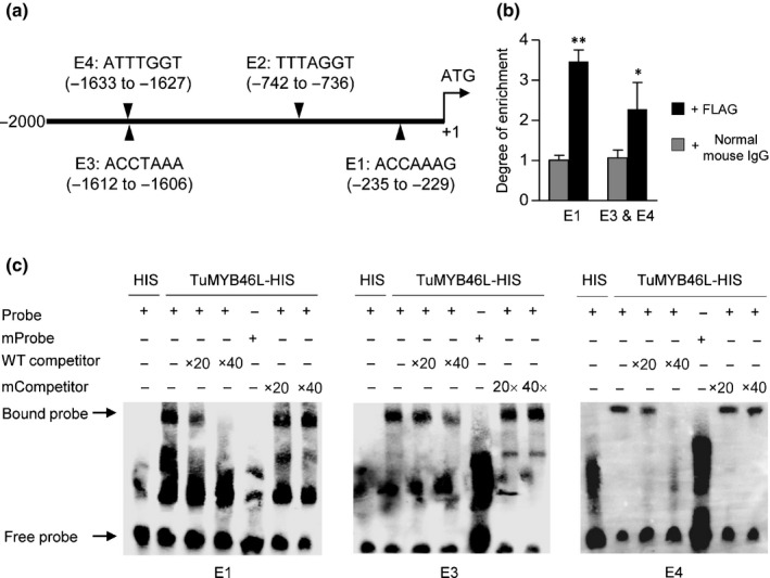
Figure 6.

Binding of TuMYB46L to TuACO3 promoter region. (a) A diagram showing the positions of four putative MYB46 binding sites (E1–E4) in the 2‐kb promoter region of TuACO3. (b) Chromatin immunoprecipitation‐quantitative (ChIP‐q)PCR assay. The assay was conducted by expressing a TuMYB46L‐FLAG protein in G1812 leaf protoplasts, with the chromatins bound to TuMYB46L‐FLAG precipitated using the FLAG antibody and subsequently quantified by qPCR. The chromatin fragments carrying the E1 element or harboring the closely spaced E3 and E4 elements were strongly enriched in the precipitation with FLAG antibody compared to the mock precipitation with normal mouse IgG. The means (± SE) each were calculated from three biological replicates, and statistically compared using Student's t‐test. *, P < 0.05; **, P < 0.01. (c) Validation of the binding of TuMYB46L to E1, E3 and E4 by EMSA. A TuMYB46L‐HIS protein expressed in Escherichia coli was purified; its binding to biotin‐labeled E1, E3 or E4 probes was tested in the absence or presence of unlabeled wild‐type (WT) probes. No specific binding was obtained with the labeled mutant probes in which the putative binding sites were all changed to AAAAAAA. Moreover, the specific binding of TuMYB46L‐HIS to E1, E3 and E4 was not affected by the presence of the unlabeled mutant probes. Three sets of independent assays were performed, which all validated the binding of TuMYB46L‐HIS to E1, E3 and E4. ACO, 1‐aminocyclopropane‐1‐carboxylic acid oxidase; MYB, myeloblastosis.
