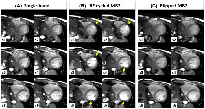
Figure 3.

Cardiac cine MB bSSFP data acquired at 3T using image‐based B 0 shimming. Single‐band (A) was acquired in 2 breath‐holds (3 slices each) and MB2 stacks in a single breath‐hold of 6 slices, using both RF cycled (B) and blipped CAIPIRINHA (C). Dark‐band artefacts (yellow arrows) are encroaching on the LV blood pool from alternate sides in the lower and upper halves of the stack with RF cycling, because of the ±FOV/4 shift in MB slices. In blipped‐CAIPI MB2 (C), the same relative slice shift is achieved without altering the bSSFP frequency response, and therefore dark‐band artefacts remain free of the heart region, comparable to single‐band case (A)
