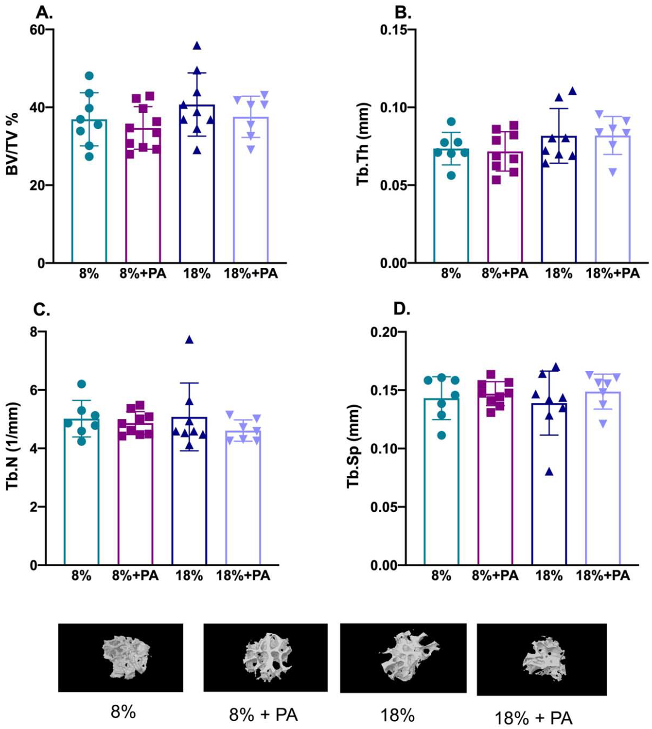
Figure 3: Bone microarchitecture was not affected by dietary PA supplementation.

Twenty-three-month-old mice were fed the indicated diets for 8 weeks and bone microarchitectural parameters of femora monitored ex vivo by μCT after sacrifice. PA did not have any significant effect on either the percentage of bone volume/total volume (BV/TV in %; Panel A); trabecular thickness (Tb.Th in mm; Panel B); trabecular number (Tb.N, per mm; Panel C); or trabecular separation (Tb.Sp, in mm; Panel D). Also shown are representative 3-dimensional μCT reconstructions of femora from mice fed the indicated diets. Data are presented as mean ± SD; n=7–10 for each diet group.
