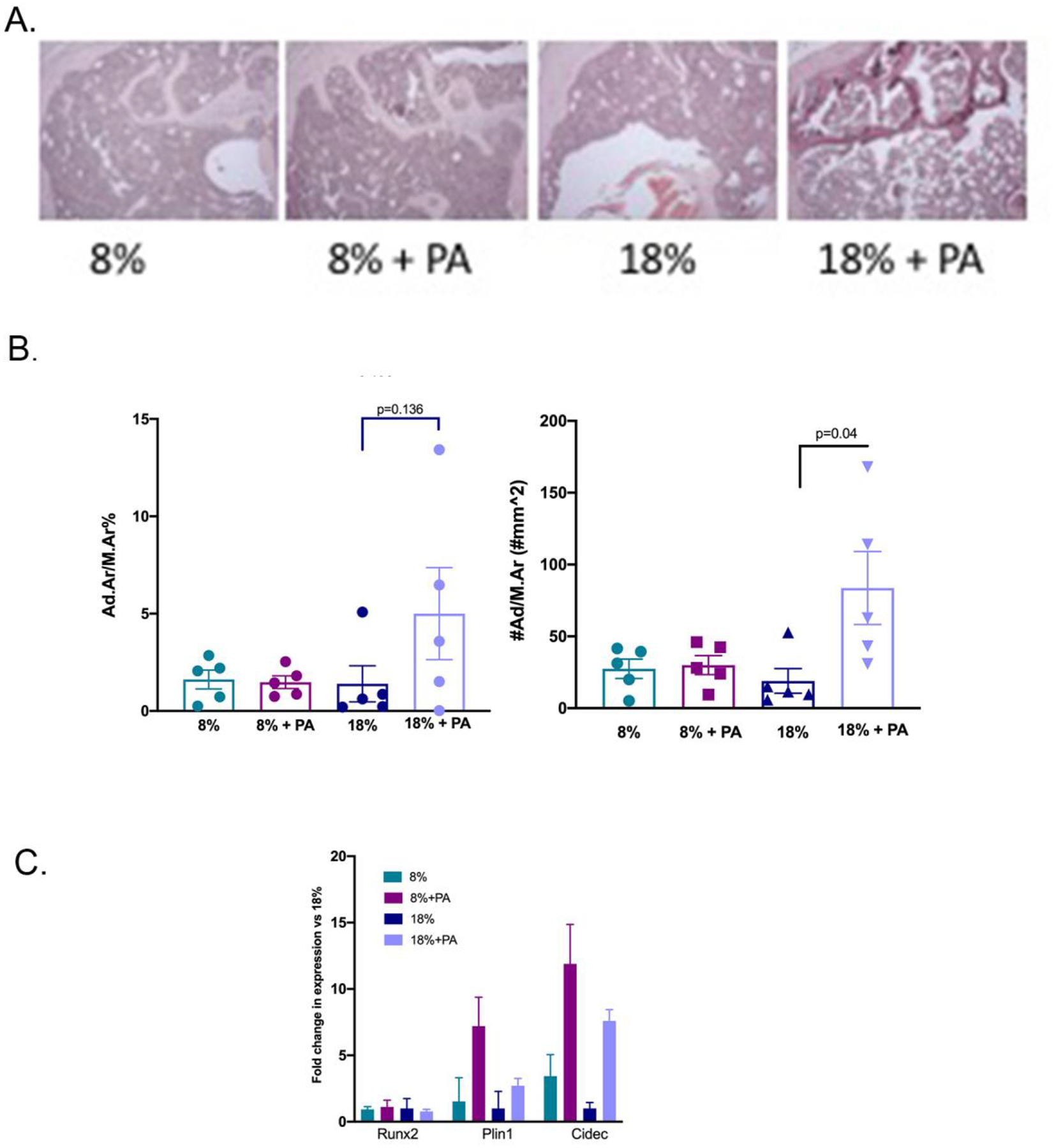
Figure 4: PA supplementation increased adipocyte number for mice on the 18% dietary protein.

Tibiae from mice fed the four diets were isolated and the bone marrow analyzed for adiposity as described in Methods. (A) Adipocyte size was calculated using adipocyte area divided by marrow area. Although there were no significant differences between the groups, adipocyte size tended to be larger for those mice on the PA and 18% dietary protein. (B) Adipocyte number/area was counted and expressed as number/mm2. Adipocyte number was significantly increased in the mice placed on the 18% protein with PA supplementation consistent with the results from Panel A. Values represent the mean ± SD of n=5 mice per group. (C) BMSCs were isolated from the humerii of animals fed for 8 weeks with the specified diets. The expression of the lipid storage genes, Plin1 and Cidec and of Runx2, associated with osteoblastic differentiation, was determined by quantitative RT-PCR as described in Methods. Values are expressed as means ± SD of n=3.
