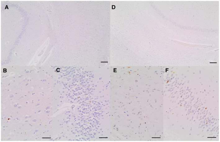
Figure 2.

A. Section of hippocampus showing dentate, molecular layer and CA1 from a case with severe dentate neuronal inclusions but minimal cell loss in CA1. B. CA1 from same slide showing a range of pathologies immunoreactive for anti‐phosphorylated TDP‐43 antibody including cytoplasmic inclusions and neurites. C. Dentate from same slide showing a range of pathologies immunoreactive for antiphosphorylated TDP‐43 antibody including cytoplasmic inclusions and neurites. D. Section of hippocampus showing dentate, molecular layer and CA1 from a case with severe dentate neuronal inclusions and severe cell loss in CA1 qualifying as HScl. E. CA1 from same slide showing a range of pathologies immunoreactive for anti‐phosphorylated TDP‐43 antibody including cytoplasmic inclusions and neurites. F. Dentate from same slide showing a range of pathologies immunoreactive for anti‐phosphorylated TDP‐43 antibody including cytoplasmic inclusions and neurites. Scale bars: A,D = 200 μm, B,C,E,F = 50 μm.
