Figure 3.

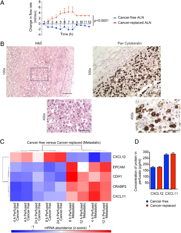
Using real‐time measurements to understand axillary lymph node (ALN) biology. (A) Cancer‐replaced ALNs (n = 3) showed a higher flow rate during perfusion than cancer‐free ALNs (n = 5) over time (two‐way ANOVA; p < 0.0001; mean with SD). (B) Cancer‐replaced ALN with pan‐cytokeratin‐positive cancer cells filling the subcapsular sinus (scale bars: 50 μm). (C) Heatmap of the five significantly differentially expressed genes between cancer‐free and cancer‐replaced (metastatic) perfused ALNs (|log2 FC| > 1 and q < 0.1; blue, down‐regulated; red, up‐regulated). (D) Concentration of CXCL12 and CXCL11 in the perfusate of cancer‐free and cancer‐replaced ALNs measured using an ELISA assay (n = 10, performed in triplicate).
