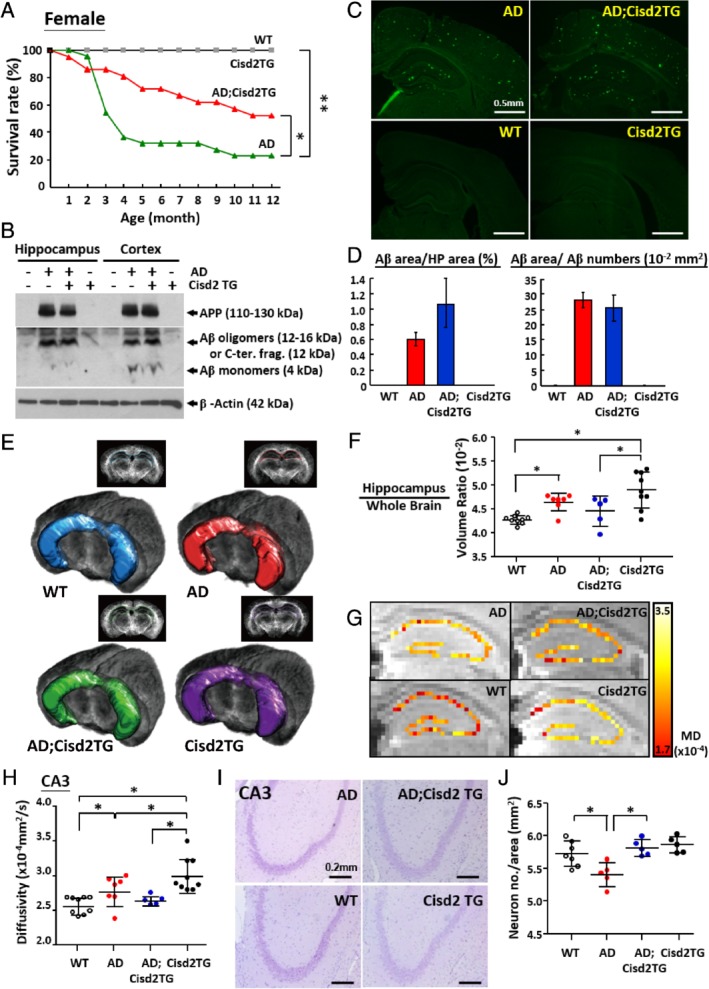
Figure 1.

Overexpression of Cisd2 prevents premature death and reduces neuronal loss in female AD mice. (A) Premature lethality among female AD mice (APPswe and PS1‐dE9 double transgenic) can be recorded as early as 1 month of age, and >60% of female AD mice died by 4 months. Two‐fold overexpression of Cisd2 in female AD mice was able to partially rescue this premature mortality and increased their survival rate. The animal numbers of each group ranged from 12 to 36 mice. (B) Western blotting analysis revealed that AD and AD;Cisd2TG mice both exhibited similar levels of precursor APP and soluble Aβ species in their hippocampus and cortex. Total protein extracts from the hippocampus and cortex of 9‐month‐old mice were separated using 15% Tricine‐SDS PAGE and detected using 6E10 antibody. (C) Thioflavin‐S staining detects extracellular Aβ plaques in WT, AD, Cisd2TG, and AD;Cisd2TG female mice. (D) Quantification of Aβ plaque number in cortex and hippocampus. Total areas of Aβ were measured using MetaMorph by setting a threshold for fluorescent intensity, and then dividing by the counting area within the hippocampus. Total areas of Aβ were divided by the number of Aβ areas in order to measure the average size of the Aβ. No significant difference in the Aβ plaque burden and average size between AD and AD;Cisd2TG mice were found. (E) Target region of interest (ROI) definitions. The ROI shows the area of hippocampus and was rendered onto the mouse brain using 2D and 3D views for the WT, AD, Cisd2TG, and AD;Cisd2TG brains. (F) Ratio of hippocampus versus whole brain volume, which was measured using TDI images. (G) DTI in the hippocampus of the WT, AD, Cisd2TG, and AD;Cisd2TG mice. (H) Quantification of mean diffusivity in the cornu ammonis 3 (CA3). (I) Nissl staining of neurons in the hippocampus (CA3) of the WT, AD, Cisd2TG, and AD;Cisd2TG mice. (J) Calculation of neuron numbers in CA3. For (C–J) mice were 12‐months‐old. Mean ± SD. *p < 0.05, **p < 0.005.
