Figure 4.

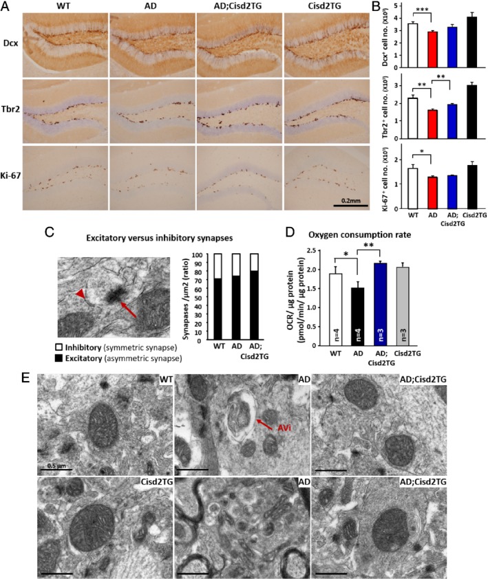
Elevated expression of Cisd2 attenuates the loss of neuronal progenitor cells and protects against Aβ‐induced mitochondrial defects. (A) Immunohistochemical staining of brain sections to label Doublecortin (Dcx), T‐box brain protein 2 (Tbr2), and antigen Ki‐67 (Ki67) in order to detect neuronal progenitor cells. (B) Quantification of cells positive for Dcx, Tbr2, and Ki‐67 in the DG region. (C) Excitatory (asymmetric) and inhibitory (symmetric) synapses revealed by TEM. Arrow indicates asymmetric synapses and arrowhead indicates symmetric synapses. Quantification results for the ratio of excitatory synapses to inhibitory synapses. (D) OCR measured using Seahorse equipment and hippocampus tissue samples obtained from the various groups of mice. (E) Ultrastructure of mitochondria in the hippocampus of the WT, AD, and AD;Cisd2 TG female mice. The mice used to create this figure were 12‐months‐old. Mean ± SD. *p < 0.05; **p < 0.005.
