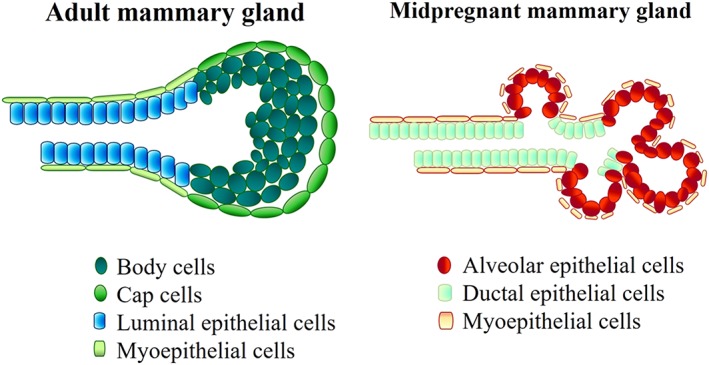
Figure 2.

Terminal end bud. On the left, adult mammary duct is characterized by a layer of cap cells (green), which surrounds body cells (dark green). Differentiated myoepithelial and luminal epithelial cells cover the inside of the duct. On the right, midpregnant mammary gland: Myoepithelial cells are on the external side of the ducts and surround the alveolar epithelial cells (red)
