Figure 1.

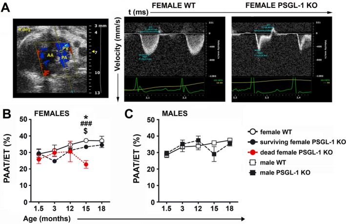
Development of pulmonary hypertension with aging in female PSGL‐1−/− mice. A, B‐mode showing the echocardiographic plane used for Doppler pulmonary flow acquisition (left), and representative Doppler pulmonary artery flow of female wild‐type (WT) mice (middle) and PSGL‐1–knockout (KO) mice (right). B, Longitudinal study of the pulmonary artery acceleration time:ejection time (PAAT:ET) ratio between 1.5 and 18 months of age in female WT mice (n = 4), surviving female PSGL‐1−/− mice (n = 6), and female PSGL‐1−/− mice that died prematurely (n = 3). C, Longitudinal study of the PAAT:ET ratio between 1.5 and 18 months of age in male WT mice (n = 6) and male PSGL‐1−/− mice (n = 6). Results are representative of 3 replicate experiments. Values are the mean ± SD. * = P < 0.05, WT versus surviving PSGL‐1−/− mice; ### = P < 0.005, WT versus dead PSGL‐1−/− mice; $ = P < 0.05, surviving PSGL‐1−/− versus dead PSGL‐1−/− mice, all by one‐way analysis of variance with Bonferroni post hoc test. AA = ascending aorta; PV = pulmonary valve; PA = pulmonary artery.
