
Figure 1.
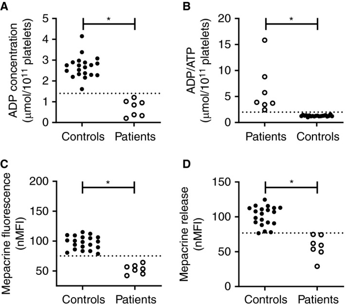
High discriminative ability of flow cytometric measurement of platelet‐dense granule content in patients with previously diagnosed δ‐SPD. (A) Platelet ADP content, expressed as µmol/1011 platelets measured with luminescence, (B) platelet ADP/ATP ratio, (C) normalized mepacrine fluorescence, and (D) mepacrine release in 20 healthy controls (closed symbols) and 7 δ‐SPD patients measured with flow cytometry. The dotted line represents the 2.5th percentile of the healthy control population. *P value < .05