Figure 3.

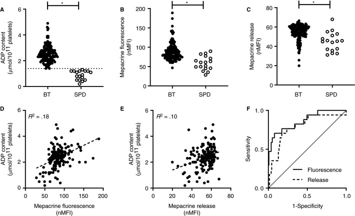
Diagnostic accuracy of flow cytometric mepacrine uptake and mepacrine release in patients with suspected platelet function disorders. (A) Platelet ADP content, expressed as µmol/1011 platelets, (B) mepacrine fluorescence expressed as normalized mean florescence intensity, (C) normalized mepacrine release for all patients included in the validation cohort. Patients were classified as bleeding tendency (BT) without δ‐SPD (BT; n = 139), or patients with a bleeding tendency and δ‐SPD (SPD; n = 17). The cutoff value for ADP content was 1.4 µmol ADP/1011 platelets. The correlation of (D) mepacrine fluorescence and (E) mepacrine release with platelet ADP content in all 156 patients with suspected platelet function disorder. (F) The discriminative ability of mepacrine fluorescence (area under the curve [AUC] 0.87) and mepacrine release (AUC 0.79) with platelet ADP as reference test plotted in a ROC curve. *P value < .05
