Figure 3.

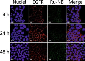
Confocal immunofluorescence microscopy images of A431 cells exposed to Ru‐NB for 4, 24, and 48 h at 37 °C showing specific binding and co‐localization of the single‐conjugated NB with EGFR. Scale bars: 20 μm.

Confocal immunofluorescence microscopy images of A431 cells exposed to Ru‐NB for 4, 24, and 48 h at 37 °C showing specific binding and co‐localization of the single‐conjugated NB with EGFR. Scale bars: 20 μm.