
Figure 1.
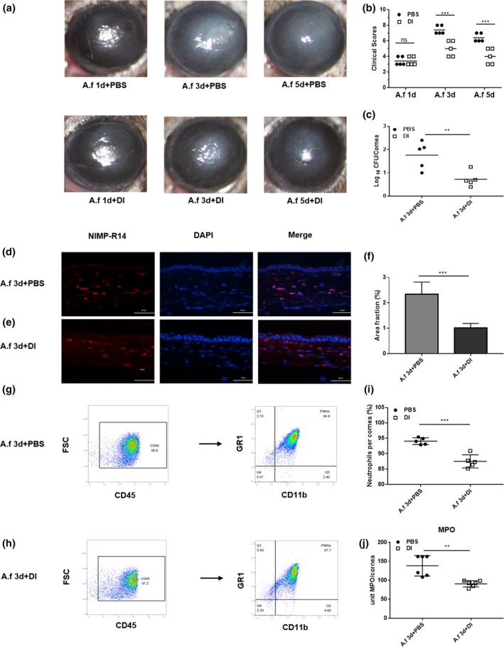
Dimethyl itaconate (DI) treatment of B6 mice. (a) Photographs of DI‐ and phosphate‐buffered saline (PBS)‐treated corneas (n = 5) taken with a slit lamp at 3 and 5 days postinfection (p.i.) showing reduced corneal opacity after DI treatment. Magnification, 25×. (b) A significant reduction in clinical scores was observed at 3 and 5 days p.i. in DI‐treated corneas (n = 5) compared with PBS‐treated corneas (n = 5). (c) The viable fungal plate count (n = 5) was reduced significantly at 3 days p.i. after DI versus PBS treatment. (d, e) Polymorphonuclear neutrophils (PMNs) were stained with NIMP‐R14, and fewer PMNs accumulated in the stroma of DI‐treated infected corneas (n = 3) than in that of PBS‐treated infected corneas (n = 3). Magnification, 400×. (f) Quantitative analysis of immunostaining images also demonstrated a reduction in PMN accumulation with DI treatment. (g–i) Flow cytometry showed that compared with PBS treatment, DI treatment decreased the percentage of PMNs in infected corneas. (j) Infected DI‐treated corneas (n = 6) showed a significantly reduced level of myeloperoxidase (MPO) at 3 days p.i. compared with PBS‐treated corneas (n = 6). The experiments were repeated two times to ensure reproducibility. P‐values are presented as the mean ± s.e.m. **P < 0.01, ***P < 0.001. A.f, Aspergillus fumigatus; DAPI, 4′,6‐diamidino‐2‐phenylindole; FSC, forward scatter; MPO, myeloperoxidase; ns, no significance.