
Figure 4.
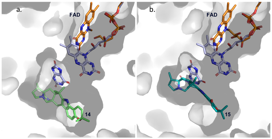
Modeled cofactor dynamics in PhqK. Flavin dynamics were modeled from HpxO crystal structure (FAD and substrate in lavender) (PDB: 3rp7) to depict the “in” conformation of the flavin relative to the substrate position. The surface representation of PhqK is shown along with the substrate and FAD from the PhqK structure (orange). Substrates 14 (a.) and 15 (b.) are shown in green and teal.