
Figure 4.
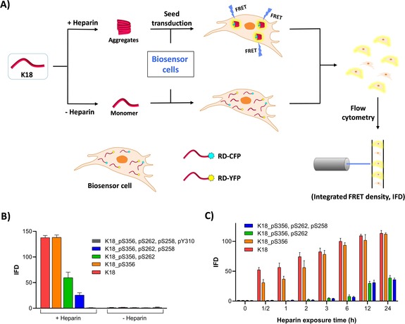
A) Schematic representation of the FRET‐based seeding assay using HEK293T biosensor cell reporter lines. Heparin‐induced aggregates are transfected into the biosensor cells using Lipofectamine. Aggregation seeded by the K18 proteins is detected as FRET emission and quantified using IFD. B) IFD measured following transduction of WT, mono‐, di‐, tri‐, and tetraphosphorylated K18 previously incubated with or without heparin for 24 h at 37 °C (N=3 repeats). C) Effect of different heparin exposure times with K18 proteins prior to transfection of the aggregates (N=3 repeats).