Fig. 1.
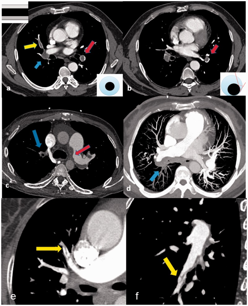
CT features of PTE: (a) acute PTE (transversal plane): filling defect of left lower pulmonary artery in “Polo mint” sign (red arrow) and filling defect of right middle pulmonary artery in “railway track” sign (yellow arrow), right lower lobe pulmonary artery occlusion (blue arrow); (b) acute PTE (transversal plane): the off-centered filling defect formed acute angles with the vessel wall (red arrow); (c) chronic PTE (transversal plane): the eccentric filling defect forming obtuse angle with the left pulmonary arterial wall (red arrow) and pouch defect of right upper pulmonary artery (blue arrow); (d) chronic PTE (maximum intensity projection): the eccentric filling defect forming obtuse angle with the right pulmonary arterial wall and pouch defect of right lower pulmonary artery (blue arrow); (e) chronic PTE (transversal plane): band-like filling defect of anterior superior lobe pulmonary artery (yellow arrow); and (f) chronic PTE (transversal plane): web-like filling defect of right lower lobe outer basal segment pulmonary artery (yellow arrow).

