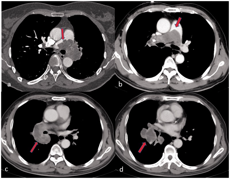
Fig. 4.
Imaging findings of PAS on CTPA (transversal plane): (a) the proximal margin of filling defect in lobular sign (red arrow); (b) a tongue sign (red arrow) of filling defect in main pulmonary artery; (c) heterogeneous enhancement (red arrow) of filling defect in right lower pulmonary artery; and (d) aneurysmal dilation and massive filling defect (red arrow) of the basal pulmonary artery is one specific finding of PAS.

