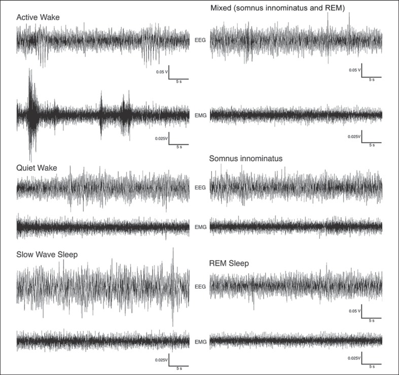
Fig. 1.
Examples of EEG and EMG polygraphs demonstrating 2-min episodes of active wake, quiet wake, SWS, SI and REM states in the rock hyrax. Waking episodes were characterized by a low-voltage, high-frequency EEG. The EMG for active waking exhibited higher voltages compared to the quiet waking state, and high-voltage spikes that likely correspond with movements were evident during this state. SWS was characterized by a high-voltage, low-frequency EEG and an EMG that was lower in amplitude than during the waking states. The EEG for both SI and REM resembled that of waking; however, during SI the EMG remained at the same amplitude as the preceding SWS episode, while during REM the EMG was reduced in amplitude.

